


Lower Urinary Tract Anatomy and Micturition Physiology Guide
Urination looks simple from the outside, but it depends on a finely timed system of muscle, nerve signalling, pressure control, and sphincter coordination. When one part fails, symptoms can range from urgency and leakage to incomplete emptying and urinary retention.
The Lower Urinary Tract includes the urinary bladder and urethra. Its main tasks are to store urine at low pressure, protect the kidneys from back-pressure, and empty at a socially appropriate time. This guide explains the core bladder anatomy, urethra function, micturition physiology, and the mechanisms behind common functional disorders.
This article is for general medical education only. It is not a substitute for assessment by a qualified clinician.

The lower urinary tract stores and empties urine as one unit
The bladder and urethra do not act as separate tubes and chambers. They form a coordinated functional system.
During storage, the bladder must relax as it fills. At the same time, the bladder outlet, urethral sphincters, and pelvic floor must maintain enough resistance to prevent leakage. During voiding, the opposite must happen. The bladder wall contracts, while the outlet and sphincters relax.
This switching behaviour is central to micturition physiology. It depends on:
The structure of the bladder wall
The tone of the bladder neck and urethra
The internal and external sphincters
The pelvic floor muscles
Sensory signals from the bladder
Autonomic and somatic nerve control
Brainstem and cortical control of timing
A useful way to understand the system is to think of it as a reservoir with a controlled outlet. The bladder stores urine without a large rise in pressure. The outlet remains closed until the nervous system permits emptying.
Bladder anatomy centres on the detrusor muscle
The urinary bladder is a hollow muscular organ in the pelvis. Its shape changes with filling. When empty, it sits low behind the pubic bone. As it fills, it rises and becomes more rounded.
The key parts of the bladder include the dome, body, base, trigone, bladder neck, and ureteric openings.
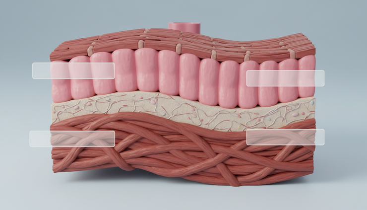
The bladder wall has several layers:
Layer | Main role |
Urothelium | Forms a specialised urine-resistant lining and helps sense stretch |
Lamina propria | Supports the lining and carries blood vessels and nerves |
Detrusor muscle | Provides the force for bladder emptying |
Adventitia or serosa | Outer covering, depending on the bladder surface |
The detrusor muscle is the main contractile layer of the bladder. It consists of smooth muscle fibres arranged in interlacing bundles rather than a single simple sheet. This arrangement allows the bladder to contract as a whole and generate pressure during voiding.
During storage, the detrusor should remain relaxed. This allows the bladder to fill without a steep rise in pressure. During voiding, detrusor contraction increases bladder pressure and drives urine through the urethra.
The urothelium was once viewed mainly as a passive barrier. It is now understood to have sensory and signalling roles. It helps detect stretch, chemical irritation, and changes in urine composition. These signals contribute to urgency, bladder sensation, and reflex control.
The trigone and bladder neck guide urine flow
The trigone is a smooth triangular area at the bladder base. Its corners are formed by the two ureteric openings and the internal urethral opening. Unlike the rest of the bladder lining, the trigone is relatively smooth and less folded.
The bladder neck is the funnel-shaped outlet between the bladder and urethra. It plays a major role in continence, especially during storage. In people with male pelvic anatomy, the bladder neck and proximal urethra also help prevent retrograde ejaculation by closing during ejaculation.
Poor bladder neck opening can contribute to obstruction and retention. Poor closure can contribute to leakage, particularly when sphincter support is weak.
Urethra function depends on structure and sphincter control
The urethra carries urine from the bladder to the outside. Its function is not only drainage. It is also a continence organ.
Urethral closure depends on several mechanisms working together:
Smooth muscle tone in the bladder neck and proximal urethra
Striated muscle control in the external urethral sphincter
Support from pelvic floor muscles and connective tissue
Mucosal sealing within the urethral lining
Proper position of the bladder neck and urethra
Urethral anatomy differs by pelvis and reproductive anatomy.
In people with female pelvic anatomy, the urethra is short and runs from the bladder neck to the external urethral opening in the vestibule. Its continence function depends strongly on pelvic floor support, urethral mucosal coaptation, and sphincter strength.
In people with male pelvic anatomy, the urethra is longer and passes through the prostate, pelvic floor, and penis. It is commonly described in sections, including the prostatic, membranous, and spongy urethra. The prostate surrounds the proximal urethra, which is why prostate enlargement can affect urine flow.
Internal and external sphincters have different roles
The internal urethral sphincter is mainly smooth muscle at the bladder neck and proximal urethra. It is under autonomic control and is not usually under conscious control.
The external urethral sphincter is striated muscle. It surrounds the urethra and is controlled by the pudendal nerve. It allows voluntary tightening, such as when a person delays urination or tries to stop urine flow.
The pelvic floor works with the external sphincter. When the pelvic floor contracts, it helps lift and compress the urethra. When voiding begins, the pelvic floor must relax so that urine can pass freely.
Micturition physiology switches between storage and emptying
The lower urinary tract has two main operating modes: storage and voiding. Each mode uses a different pattern of nerve activity.
Storage is controlled mainly by sympathetic and somatic activity
As the bladder fills, stretch receptors send sensory signals through pelvic nerves to the spinal cord and brain. At low to moderate volumes, the system favours storage.
During storage:
The detrusor relaxes
The bladder neck and proximal urethra contract
The external sphincter remains active
Bladder pressure stays low
Urethral pressure stays higher than bladder pressure
Sympathetic nerves, mainly through the hypogastric nerve, support storage. Beta-adrenergic activity relaxes the detrusor. Alpha-adrenergic activity contracts the bladder neck and proximal urethra.
Somatic nerves, mainly through the pudendal nerve, keep the external sphincter active. This guarding reflex increases sphincter activity when bladder pressure rises, such as during coughing, lifting, or sudden movement.
Voiding is controlled mainly by parasympathetic activity
When bladder filling reaches a suitable threshold, sensory signals become stronger. The brain then decides whether voiding is appropriate. If it is not, cortical control suppresses the voiding reflex.
When voiding is permitted, the pontine micturition centre coordinates the change from storage to emptying.
During voiding:
The pelvic floor relaxes
The external sphincter relaxes
The bladder neck opens
Parasympathetic nerves stimulate detrusor contraction
Urine flows through a low-resistance urethra
Parasympathetic fibres travel mainly through the pelvic nerves from the sacral spinal cord. They act on muscarinic receptors, especially M3 receptors, in the detrusor muscle. This produces coordinated detrusor contraction.
Normal urination requires more than a strong bladder contraction. It requires the outlet to relax at the same time.
If the detrusor contracts against a closed or poorly relaxed outlet, pressure rises and flow falls. If the outlet opens but the detrusor is weak, emptying may be slow or incomplete.

Functional disorders often reflect a breakdown in coordination
Many urinary symptoms make sense when viewed through anatomy and physiology. A symptom is rarely just a bladder problem or just a urethral problem. It often reflects altered pressure, nerve control, outlet resistance, or muscle support.
Overactive bladder involves urgency and detrusor overactivity
Overactive bladder is a symptom syndrome associated with urgency, usually with frequency and nocturia. Some people also have urgency urinary incontinence.
In urodynamic testing, some patients show detrusor overactivity. This means the detrusor contracts involuntarily during bladder filling. These contractions can create sudden urgency and leakage if bladder pressure exceeds urethral closure pressure.
Common contributing mechanisms include increased bladder sensory signalling, altered central inhibition, detrusor instability, and irritation from infection, stones, or inflammation. Neurological conditions can also affect the pathways that suppress reflex bladder contractions.
Urinary retention can result from weak contraction or high outlet resistance
Urinary retention occurs when the bladder does not empty properly. It may be acute, with sudden painful inability to pass urine, or chronic, with incomplete emptying and raised post-void residual urine.
Broad mechanisms include:
Weak or underactive detrusor contraction
Obstruction at the bladder neck or urethra
Poor sphincter relaxation during voiding
Medication effects
Neurological impairment
Post-operative urinary retention
In people with male pelvic anatomy, prostate enlargement is a common cause of increased outlet resistance. Urethral stricture, pelvic organ prolapse, constipation, and pelvic floor dysfunction can also affect emptying.
Chronic retention can be clinically quiet. Some people mainly notice weak flow, hesitancy, straining, repeated voiding, or a feeling of incomplete emptying.
Stress urinary incontinence reflects outlet support failure
Stress urinary incontinence is leakage during raised abdominal pressure, such as coughing, laughing, running, or lifting. The key issue is not an urgent bladder contraction. It is a failure of urethral closure pressure to stay above bladder pressure.
This may happen because of:
Reduced pelvic floor support
Urethral hypermobility
Intrinsic sphincter weakness
Effects of childbirth, pelvic surgery, ageing, or hormonal change
Neurological or connective tissue factors
The distinction matters. Urgency incontinence and stress incontinence can feel similar to the person experiencing leakage, but the mechanism and treatment approach can differ.
Neurogenic bladder depends on lesion level and pathway disruption
The bladder depends on communication between the brain, spinal cord, peripheral nerves, detrusor, and sphincters. Neurological disease or injury can disrupt this system at different levels.
Some lesions cause detrusor overactivity. Others cause poor detrusor contraction. Some cause detrusor sphincter dyssynergia, where the bladder contracts but the external sphincter does not relax properly.
This can create high bladder pressures and incomplete emptying. In some cases, protecting kidney function becomes a major priority.
Urodynamics measures pressure, flow, and coordination
Urodynamic testing helps explain symptoms by measuring lower urinary tract function. It is not needed for every urinary complaint, but it can be useful when symptoms are complex, treatment has failed, surgery is being considered, or neurological disease is present.
Common assessments include:
Test | What it helps assess |
Uroflowmetry | Urine flow rate and voiding pattern |
Post-void residual measurement | How much urine remains after voiding |
Filling cystometry | Bladder sensation, capacity, compliance, and involuntary contractions |
Pressure-flow study | Detrusor pressure and outlet resistance during voiding |
Electromyography | External sphincter or pelvic floor activity in selected cases |
Good interpretation links the test result with the symptom story. A low flow rate may reflect obstruction, weak detrusor contraction, or both. Leakage during filling may arise from detrusor overactivity, sphincter weakness, or raised abdominal pressure.
This is why anatomy matters. The diagnosis becomes clearer when pressure, flow, muscle activity, and clinical history all point in the same direction.
Why this anatomy matters in clinical care
Treatment choices often target a specific point in the storage or voiding pathway.
For urgency and overactive bladder, care may include bladder training, fluid and caffeine adjustment, pelvic floor therapy, antimuscarinic medicines, beta-3 agonists, botulinum toxin injections, neuromodulation, or other specialist options.
For stress urinary incontinence, pelvic floor muscle training often forms the foundation. Depending on severity and cause, pessaries, urethral bulking, sling surgery, or other procedures may be considered.
For retention, management depends on cause and risk. Options may include treating obstruction, reviewing medicines, intermittent catheterisation, pelvic floor relaxation work, or managing neurological disease.
For recurrent symptoms, male reproductive or pelvic health concerns, or urinary problems affecting quality of life, a specialist review can help connect symptoms with anatomy and physiology. If you are seeking assessment in the UK, you can book an appointment with The Male Factor.
Frequently asked questions
What is the main function of the detrusor muscle?
The detrusor muscle contracts to empty the bladder. During storage, it should stay relaxed so the bladder can fill at low pressure.
What does the urethra do besides carry urine?
The urethra also helps maintain continence. Its sphincters, lining, smooth muscle tone, and pelvic floor support keep urine in the bladder during storage.
What is the micturition reflex?
The micturition reflex is the nerve-controlled process that switches the bladder from storage to emptying. It involves detrusor contraction, sphincter relaxation, and coordination between the bladder, spinal cord, brainstem, and brain.
Why can urinary incontinence have different causes?
Leakage can happen because the bladder contracts too soon, the urethra does not close well enough, or both. This is why urgency incontinence and stress incontinence need different assessment and treatment plans.
When are urodynamic tests useful?
Urodynamics may help when symptoms are unclear, severe, recurrent, neurological, or being assessed before surgery. The tests show how bladder pressure, urine flow, and sphincter function behave during filling and voiding.

The key takeaway
The bladder and urethra are best understood as a coordinated pressure system. Storage depends on a relaxed detrusor and a closed outlet. Emptying depends on detrusor contraction with sphincter and pelvic floor relaxation.
When this timing fails, symptoms such as urgency, urinary incontinence, weak flow, and urinary retention can develop. A clear grasp of bladder anatomy, urethra function, and micturition physiology gives clinicians the framework to diagnose the problem and choose treatment that matches the underlying mechanism.


Comments